
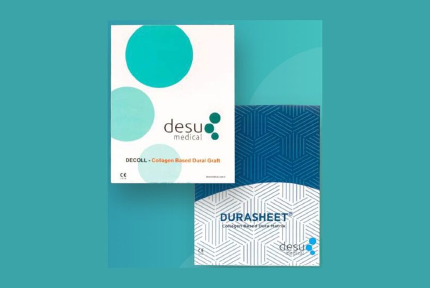
Collagen graft resorption is the quiet endpoint that separates a biological dural repair from a mechanical one. A permanent membrane holds the defect closed indefinitely. A resorbable one disappears, leaving the patient’s own tissue in its place. Desu Medical manufactures dural grafts from pure Type I bovine collagen because that second outcome is the better one.
The Sequence After Placement
Resorption is not a single event. It is a sequence that overlaps with healing.
Immediate Barrier Phase
From the moment of placement, the membrane acts as a physical barrier. DECOLL is produced with an impermeable pore structure that resists cerebrospinal fluid transit. This phase carries the repair while biology gets to work.
Cellular Infiltration
Fibroblasts migrate inward from the dural margins. Because the scaffold is Type I collagen, the same protein those cells normally encounter, integrin-mediated attachment proceeds without a preparatory inflammatory phase. The cells then begin depositing new extracellular matrix within and around the scaffold.
Enzymatic Breakdown
Endogenous collagenases progressively degrade the implanted matrix, exactly as they process native collagen turnover. Consequently, the scaffold thins as host tissue thickens.
Tissue Replacement
Finally, the repair site consists of autologous connective tissue continuous with the surrounding dura. The graft, functionally and structurally, is gone.
Why Purity Governs the Timeline
Residual non-collagenous proteins, lipids and cellular debris provoke reaction, and reaction distorts remodelling. Encapsulation, chronic inflammation and fibrotic walling-off all interfere with the orderly sequence above. Therefore purification to pure Type I collagen is not simply a safety measure. It is what allows collagen graft resorption and tissue deposition to stay in balance.
In addition, Desu’s process preserves the triple-helical structure rather than heavily crosslinking it. Aggressive crosslinking extends dwell time but changes the material’s biological signature and slows the handover to host tissue.
Clinical Consequences
- No removal procedure. The graft does not require a secondary operation for retrieval or revision.
- Simpler reoperation. Patients returning for cranioplasty or further decompression encounter host tissue rather than a permanent membrane.
- Cleaner imaging. No residual implant remains at the repair site to complicate interpretation.
- Lower long-term interface risk. A material that is no longer present cannot generate a chronic response.
Factors That Influence Resorption
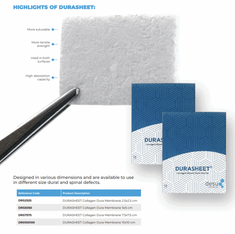
Timelines vary with defect size, local vascularity, patient age and comorbidity. Larger reconstructions naturally take longer to populate than focal repairs. Furthermore, the surgical technique matters: generous overlap onto well-vascularised dura gives cells a shorter distance to travel. For this reason the DECOLL range spans 2.5 to 10 cm squares, referenced DCL2525 through DCL100100, so the surgeon can size for overlap rather than economy.
Evidence and Testing
Absorption behaviour and tissue integration have been evaluated as part of the Desu technical documentation, alongside mechanical durability. Biocompatibility testing follows the ISO 10993 series, and manufacturing operates under an ISO 13485 quality management system with CE certification. Institutions can request the technical file from [email protected].
Frequently Asked Questions
How long does resorption take?
It depends on defect size, vascularity and patient factors. The membrane provides barrier function throughout the healing window and is then replaced by host tissue.
Does resorption weaken the repair before healing completes?
The processes overlap by design. Host matrix deposition proceeds alongside scaffold breakdown rather than after it.
Is the same behaviour expected from DURASHEET?
Yes. DURASHEET shares the same pure Type I bovine collagen specification and is likewise resorbable.

Resorbable Versus Permanent: The Long View
The difference between a resorbable and a permanent dural implant is not visible in the first week. It becomes visible at reoperation, at follow-up imaging, and in the small proportion of patients who develop a chronic response to a retained material. A membrane that has been replaced by host tissue cannot produce any of these.
For younger patients in particular, the time horizon matters. A permanent implant placed at thirty is present at seventy. A resorbable collagen scaffold placed at thirty is gone within months, replaced by the patient’s own connective tissue.
Surgical Factors That Support Remodelling
- Overlap onto vascularised dura. Cells enter from the margins, so healthy margins matter more than graft area.
- Avoid tension. A graft under tension apposes poorly, and poor apposition slows integration.
- Debride devitalised edges. Non-viable tissue contributes no cells to the repair.
- Handle minimally. The membrane is ready to use, so extended manipulation adds nothing.
What Resorption Does Not Mean
Resorbable does not mean fragile, and it does not mean the barrier disappears before healing completes. The processes are designed to overlap: host matrix deposition proceeds alongside scaffold breakdown rather than after it. Equally, resorption is not a substitute for adequate surgical technique. A poorly apposed graft does not become a good repair through biology alone.
Explaining Resorption to Patients and Colleagues
Patients frequently ask whether something permanent has been left inside them. The answer with a purified collagen graft is straightforward: the material is a temporary scaffold made of the same protein already present throughout the body, and it is gradually replaced by the patient’s own tissue. Nothing remains to be removed later.
The same explanation is useful with colleagues in radiology and with theatre staff. Radiologists interpreting follow-up imaging benefit from knowing that no permanent membrane is present at the repair site. Theatre teams benefit from understanding why the material is handled minimally and placed without preparation. Our clinical team provides product education covering these points for departments adopting the range.
One further point is worth making to procurement colleagues. Resorption removes a category of long-term risk from the cost model entirely, because a material that is no longer present cannot generate a late complication, cannot require removal, and cannot complicate a future operation at the same site. That is difficult to price in a tender spreadsheet, yet it is one of the clearest advantages a purified collagen scaffold holds over a permanent implant.
Request the Technical Documentation
Detailed specifications for both membranes, including absorption and integration data summaries, are available through desu.tr/. To obtain the full technical file, arrange evaluation samples or request a quotation, contact [email protected].
Information for healthcare professionals. Refer to the instructions for use.
About Desu Medical
Desu Medical is a medical research and development company founded in 2007 and headquartered in Ankara, Turkiye. The company designs and manufactures neurosurgical devices in-house, including hydrocephalus shunt systems, external ventricular drainage sets, catheters, intracranial pressure monitoring, bone grafts and the collagen dural graft range comprising DECOLL and DURASHEET. Every dural graft is produced from pure Type I bovine collagen sourced from controlled and traceable tissue, gamma sterilised, and supplied ready to use.
Manufacturing operates under an ISO 13485 certified quality management system, the dural graft range carries CE certification, and biocompatibility has been evaluated across the ISO 10993 series. Desu supplies healthcare professionals internationally through direct institutional relationships and distribution partners. For product specifications, CE documentation, evaluation samples or quotations, visit desu.tr/ or contact the team at [email protected].
Frequently Asked Questions
What is collagen graft resorption?
Collagen graft resorption is the gradual breakdown of the collagen scaffold as the patient’s own cells migrate in and lay down new tissue, until no permanent membrane remains.
How long does collagen graft resorption take?
The timeline of collagen graft resorption varies with defect size, local vascularity, patient age and comorbidity.
Does purity affect collagen graft resorption?
Yes. With pure Type I bovine collagen, collagen graft resorption proceeds as remodelling into host tissue rather than as a reaction to residual impurities.
