
Chiari duraplasty is a common step in the surgical treatment of Chiari malformation type I. After the bone at the back of the skull and the first cervical vertebra has been removed, the surgeon opens the dura mater and sews in a patch to enlarge the space at the craniocervical junction. The choice of graft and the quality of the closure have a direct influence on how well the decompression works and on the risk of cerebrospinal fluid (CSF) complications. This article reviews the reasons for Chiari duraplasty, the graft options and the technical points that matter.
Chiari Malformation Type I in Brief
In Chiari malformation type I, the lower part of the cerebellum, the cerebellar tonsils, extends below the foramen magnum into the upper spinal canal. This crowding can obstruct the normal flow of CSF between the head and the spine. Patients may present with occipital headaches that worsen with coughing or straining, neck pain, balance problems or symptoms of syringomyelia, a fluid-filled cavity within the spinal cord. Many people with tonsillar descent on MRI have no symptoms and do not need surgery.
Surgical Options: With or Without Duraplasty
The goal of surgery is to restore CSF flow at the craniocervical junction and relieve pressure on the brainstem and spinal cord. Two main approaches are used:
- Posterior fossa decompression without duraplasty: removal of bone, sometimes with splitting of the outer dural layer, leaving the dura closed.
- Posterior fossa decompression with duraplasty: the dura is opened and enlarged with a graft, which increases the volume available for the cerebellar tonsils and CSF.
The comparative merits of these approaches continue to be studied. Chiari duraplasty can provide a more complete decompression, especially when a syrinx is present, while bone-only decompression avoids opening the dura and the CSF-related complications that can follow. The decision depends on the patient’s anatomy, symptoms, imaging and the surgeon’s experience.
Why the Graft Matters in Chiari Duraplasty
Once the dura is opened, the graft becomes part of the boundary of the subarachnoid space. Problems that have been reported after Chiari duraplasty include pseudomeningocele, CSF leakage through the wound, aseptic (chemical) meningitis and adhesions that can re-obstruct CSF flow. Graft material, graft size and closure technique all influence these risks. Our article on pseudomeningocele covers that complication in detail.
Graft Options for Chiari Duraplasty
- Autologous pericranium: harvested through the same incision, no added cost and no foreign material, but it requires additional dissection and its size is limited.
- Collagen grafts of animal origin: readily available in several sizes, easy to trim and designed to be remodelled into host tissue over time.
- Other biological grafts: such as processed pericardium.
- Synthetic grafts: made from manufactured polymers, some of which remain permanently.
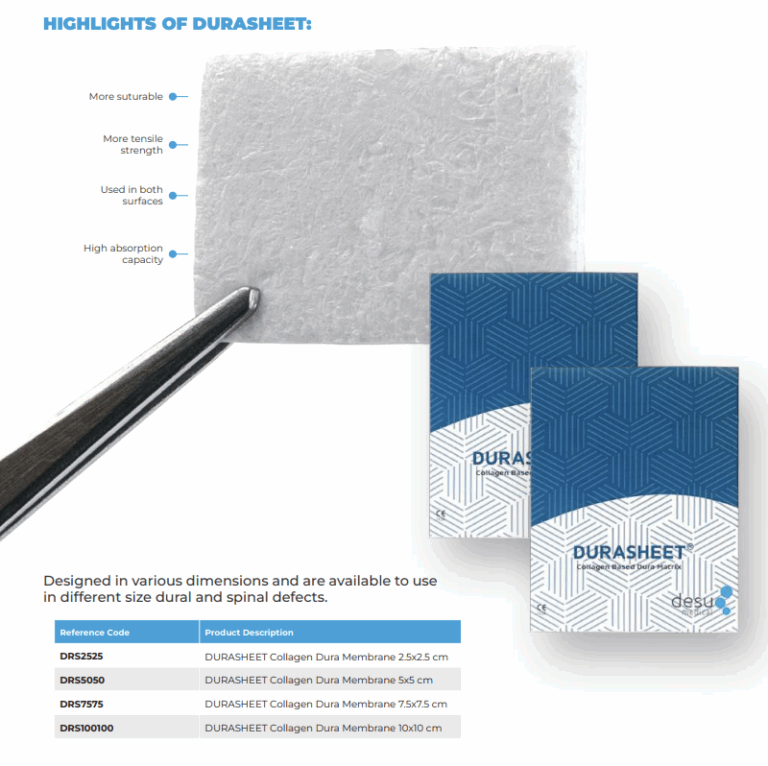
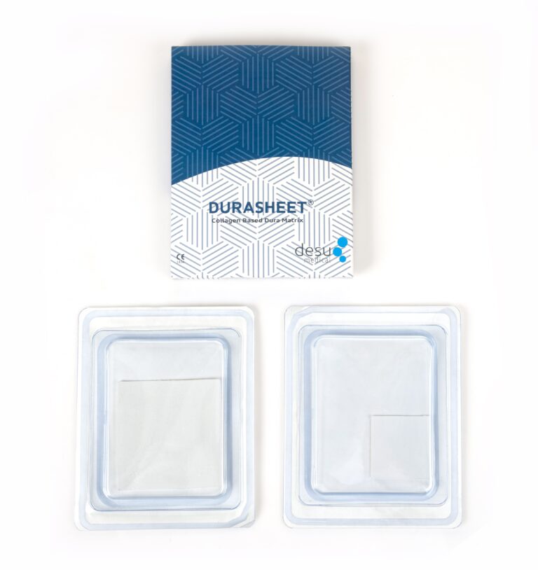
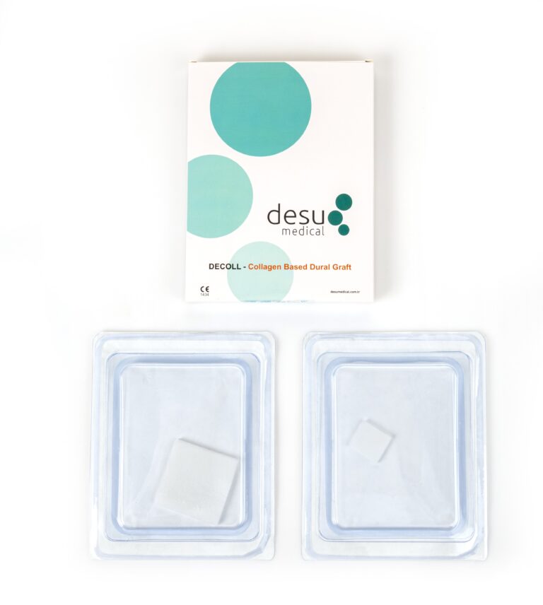
Natural collagen grafts made from purified Type I bovine collagen are a frequent choice because they combine availability with biological integration. Desu’s DECOLL and DURASHEET membranes are made from natural, pure Type I bovine collagen, not synthetic polymers.
Technical Points in Chiari Duraplasty
Durotomy and Arachnoid
Many surgeons use a Y-shaped durotomy that extends from the posterior fossa down over the upper cervical spine. Whether to open the arachnoid and manipulate the tonsils is a matter of surgical judgement. Preserving the arachnoid where possible keeps a natural barrier between the CSF and the graft.
Sizing the Patch
The patch should be large enough to create a genuinely expanded space without excessive folds. A graft that is trimmed to the shape of the defect, with an overlap at the edges, is easier to close watertight. DECOLL is available in square formats from 2.5 x 2.5 cm to 10 x 10 cm and can be cut with surgical scissors to the required shape.
Watertight Closure
The graft is sutured to the dural edges with a running or interrupted technique. Many teams test the closure with a Valsalva manoeuvre performed by the anaesthetist and reinforce small leaks with additional sutures or an adjunct. Layered closure of muscle, fascia and skin then supports the dural repair. Our guide to CSF leak prevention describes these steps.
Postoperative Considerations
After Chiari duraplasty, patients are observed for wound swelling, CSF leakage, fever and signs of meningitis. Headache and neck pain are common in the first days. MRI may be used later to confirm decompression and, where present, a reduction in the syrinx. Any persistent fluid collection or wound leak should be assessed promptly by the surgical team.
Summary
Chiari duraplasty can provide an effective decompression of the craniocervical junction, and the dural graft plays a central role in that result. A graft that is easy to size, conforms to the anatomy, supports a watertight closure and integrates into host tissue helps the surgeon achieve a reliable repair.
Frequently Asked Questions
Is duraplasty always needed in Chiari surgery?
No. Some patients are treated with bone decompression alone. Whether Chiari duraplasty is performed depends on the anatomy, symptoms, the presence of a syrinx and the surgeon’s judgement.
Which graft materials are used for Chiari duraplasty?
Options include autologous pericranium, collagen grafts of animal origin, other biological grafts and synthetic materials. Natural Type I collagen grafts are widely used because they are available in several sizes and integrate into host tissue.
What complications can follow Chiari duraplasty?
Reported complications include pseudomeningocele, CSF leakage, aseptic meningitis and wound problems. Careful sizing, watertight closure and layered wound closure help reduce these risks.
References and Further Reading
- PubMed: Chiari duraplasty graft
- PubMed: posterior fossa decompression with versus without duraplasty
- Desu Medical, DECOLL collagen dura membrane
About this article
Prepared by the Desu Medical editorial team. Desu Medical has designed and manufactured neurosurgical devices in Ankara, Türkiye, since 2007, including the DURASHEET and DECOLL collagen dura membranes, which are made from natural, pure Type I bovine collagen. Manufacturing operates under an ISO 13485 quality management system and the dural graft range is CE certified. This article is written for healthcare professionals and general information; it does not replace the Instructions for Use or clinical judgement. Last reviewed: October 2026.

1 Comment
Posterior Fossa Dural Closure in 5 Effective Steps
[…] may be swollen. An expansile duraplasty gives room to the posterior fossa contents; our article on Chiari duraplasty describes this […]