
A pseudomeningocele is a collection of cerebrospinal fluid (CSF) that has escaped through a defect in the dura and gathered in the soft tissues outside it. Unlike a true meningocele, it is not lined by dura or arachnoid, but by a capsule of scar tissue that forms around the fluid. It can occur after spinal surgery, after posterior fossa operations and, less often, after supratentorial craniotomy. This article explains why it develops, how it is recognised and how dural grafts are used when repair is required.
How a Pseudomeningocele Forms
Every pseudomeningocele starts with a breach in the dura through which CSF can pass. The breach may be an incidental durotomy during spine surgery, a dural opening that was not fully closed, a suture line that leaked under pressure, or a graft that did not seal the defect. CSF tracks into the surgical bed, and if the overlying muscle and fascia are closed well enough to stop it reaching the skin, it collects as a fluid pocket. Over time the body forms a fibrous capsule around it.
Risk Factors
- Unrecognised or difficult durotomy during lumbar or cervical spine surgery.
- Posterior fossa surgery, including Chiari decompression, where CSF pulsation is high and soft tissue closure can be challenging.
- Raised CSF pressure from hydrocephalus or idiopathic intracranial hypertension.
- Revision surgery and previous radiotherapy, which leave tissue scarred and less able to hold sutures.
- Poor soft tissue closure, with dead space left above the dura.
Symptoms and Signs
Many small collections cause no symptoms and are found incidentally on imaging. When symptoms occur, the most common is a soft, sometimes fluctuant swelling under the wound that may enlarge when the patient coughs or strains. Some patients have positional headache, especially if CSF loss is significant. After spinal surgery, nerve roots can herniate into the collection and cause radicular pain. Clear fluid leaking from the wound indicates a CSF fistula, which carries a higher risk of infection and needs prompt attention.
Diagnosis
Magnetic resonance imaging is the main tool for diagnosing a pseudomeningocele. It shows the size of the collection, its relationship to the dura and, in some cases, the tract leading to the dural defect. Ultrasound can be useful for superficial collections, particularly in children. If a CSF leak through the wound is suspected, testing the fluid for beta-2 transferrin helps confirm that it is CSF.
Conservative Management
Small, asymptomatic collections are often observed, since many shrink over time as the defect seals. Measures may include a compressive dressing, avoidance of straining and, in selected patients, a short period of lumbar CSF drainage to reduce pressure while the dura heals. Any underlying hydrocephalus should be identified and treated, because a pseudomeningocele will not settle while CSF pressure remains high.
Surgical Repair of a Pseudomeningocele
Surgery is considered for a pseudomeningocele that is large, enlarging, symptomatic or associated with a CSF fistula through the skin. The principles of repair are:
- Explore the wound and drain the collection.
- Find the dural defect, which may be small and hidden among scar tissue.
- Close the defect by direct suture if the edges allow it, or with a dural graft when they do not.
- Reinforce the repair with an onlay graft or sealant where appropriate.
- Close the soft tissues in layers, eliminating dead space so that fluid cannot collect again.
The Role of Dural Grafts
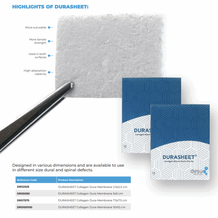
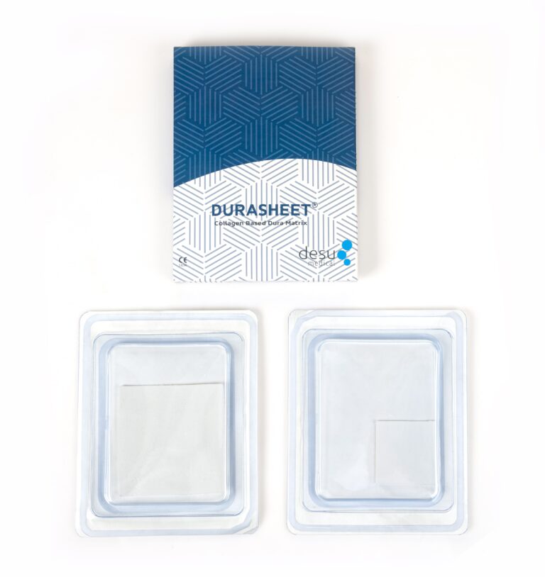
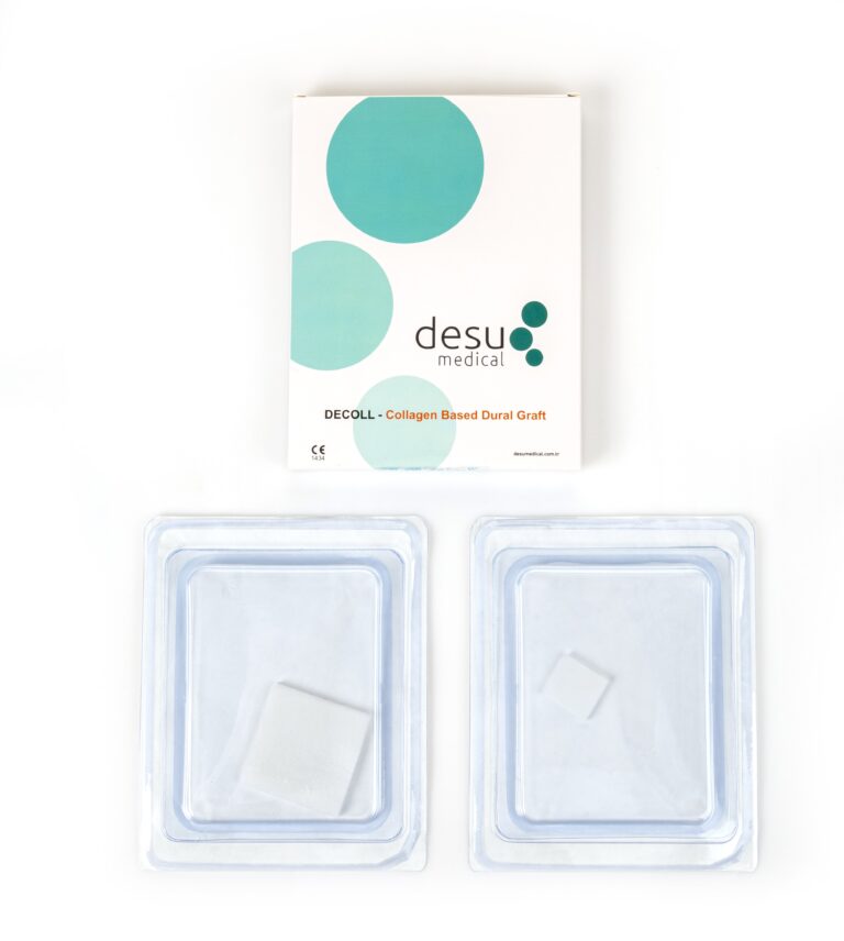
When the dural edges are retracted, thinned or scarred, a graft is needed to restore a continuous barrier. Collagen grafts are often chosen because they are soft enough to conform to an irregular defect and can be cut to the exact shape required. Desu’s DURASHEET and DECOLL membranes are made from natural, pure Type I bovine collagen with an impermeable pore structure that acts as a barrier against CSF, and they are gradually replaced by the patient’s own connective tissue. They can be sutured or used as an onlay according to the surgeon’s technique and the Instructions for Use.
Recurrence After Repair
A repaired pseudomeningocele can recur if the dural defect was not fully closed, if dead space remained in the soft tissues or if CSF pressure stays high. For this reason, surgeons check the repair under Valsalva before closing, and investigate for hydrocephalus or raised intracranial pressure when a collection returns.
Prevention Is Better Than Repair
The most effective way to deal with a pseudomeningocele is to prevent it. Careful dural closure at the first operation, testing the repair with a Valsalva manoeuvre, using a graft when the dura cannot be closed primarily without tension, and a meticulous multilayer soft tissue closure all reduce the risk. Our guide to watertight dural closure and our article on spinal durotomy repair discuss these techniques in detail.
Summary
A pseudomeningocele results from CSF escaping through a dural defect into the surrounding tissue. Many settle without surgery, but large, symptomatic or leaking collections require exploration, closure of the dural defect, often with a dural graft, and careful soft tissue reconstruction.
Frequently Asked Questions
Is a pseudomeningocele dangerous?
Many are small and harmless and resolve on their own. A pseudomeningocele becomes a concern when it grows, causes symptoms, compresses nerves or leaks through the skin, because a leak increases the risk of infection.
Does every pseudomeningocele need surgery?
No. Small collections without symptoms are often observed. Surgery is considered for large, enlarging or symptomatic collections, or when there is a CSF leak through the wound.
Why is a dural graft used in repair?
If the edges of the dural defect cannot be brought together without tension, a graft restores a continuous barrier so that CSF can no longer escape.
References and Further Reading
- PubMed: pseudomeningocele management
- PubMed: incidental durotomy and pseudomeningocele
- Desu Medical, DURASHEET collagen dura membrane
About this article
Prepared by the Desu Medical editorial team. Desu Medical has designed and manufactured neurosurgical devices in Ankara, Türkiye, since 2007, including the DURASHEET and DECOLL collagen dura membranes, which are made from natural, pure Type I bovine collagen. Manufacturing operates under an ISO 13485 quality management system and the dural graft range is CE certified. This article is written for healthcare professionals and general information; it does not replace the Instructions for Use or clinical judgement. Last reviewed: October 2026.
