
Infection after cranial or spinal surgery is uncommon, but when it involves the dural repair it can be serious, leading to meningitis, wound breakdown, graft removal and further operations. Preventing a duraplasty infection depends on many small measures taken before, during and after surgery. This article summarises those measures for surgeons, theatre teams and ward staff. It is general information and does not replace local infection control policies.
Why Duraplasty Infection Matters
Any implant placed in the body can become a surface on which bacteria settle. When a dural graft is used, the infection may involve the graft, the epidural space, the bone flap or the subdural space. Because the dura is the barrier that protects the brain and spinal cord, an infected repair can allow infection to reach the cerebrospinal fluid (CSF). A duraplasty infection often requires surgical washout and removal of infected material, as well as a prolonged course of antibiotics.
Risk Factors for Duraplasty Infection
- CSF leakage: a leak through the wound creates a direct path for bacteria to reach the CSF.
- Long operations and repeated opening of the wound.
- Revision surgery and previous radiotherapy.
- Patient factors: diabetes, smoking, poor nutrition, obesity and immunosuppression.
- Wounds near the frontal sinus or communicating with the paranasal sinuses after skull base surgery.
- External drains left in place for a long time.
Phase 1: Before Surgery
Lowering the risk of duraplasty infection starts before the patient reaches theatre. Preparation begins on the ward. Patients should be screened and optimised where possible: blood glucose control in diabetics, advice on stopping smoking, and treatment of existing skin or urinary infections. Hair removal, if needed, is performed with clippers rather than razors, close to the time of surgery. Many hospitals follow national guidance on preoperative bathing and nasal decolonisation for selected patients.
Antibiotic prophylaxis is given according to local protocol, usually within the hour before the incision, so that tissue levels are adequate when the skin is opened. The choice of drug and the need for repeat doses during long operations are set by local guidelines.
Phase 2: During Surgery
Theatre Discipline
Limiting the number of people entering and leaving the theatre, keeping doors closed and minimising unnecessary movement all reduce airborne contamination. Skin preparation with an appropriate antiseptic and careful draping follow standard practice.
Graft Handling
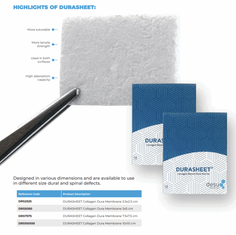
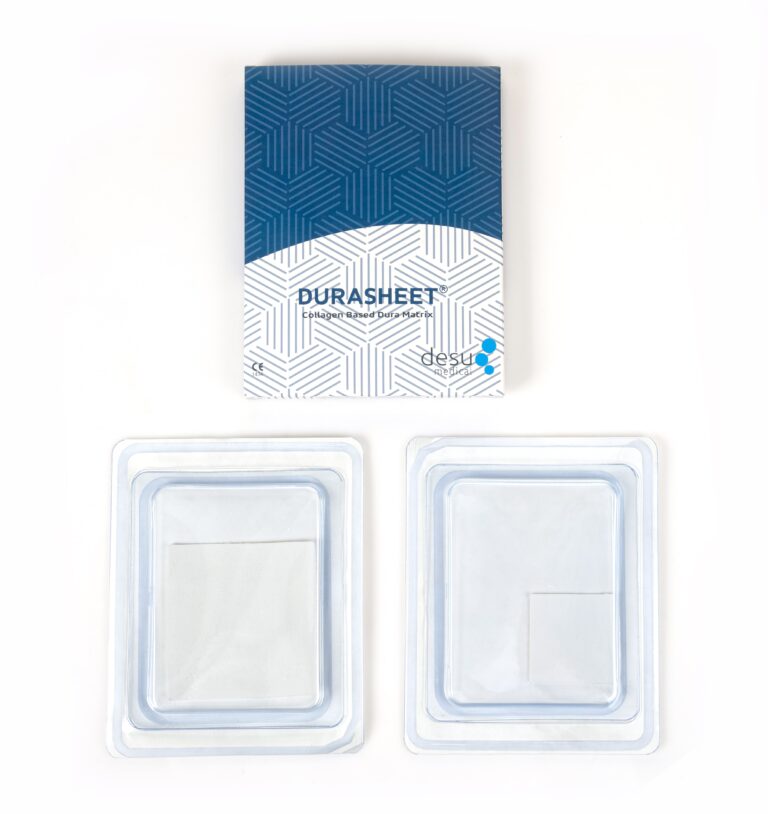
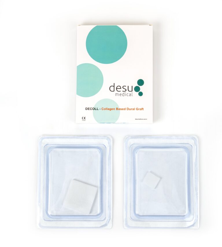
The graft should stay in its sterile package until it is needed, be handled with clean instruments and be placed promptly. Opened but unused grafts must be discarded, never re-sterilised. Our guide to dural graft handling describes these steps in detail. Desu’s DURASHEET and DECOLL collagen dura membranes are gamma sterilised, single-use and ready to use without pre-treatment, which limits handling time before placement.
A Watertight Closure
The single most important surgical measure against duraplasty infection is preventing CSF leakage. A careful dural closure, a graft where primary closure is not possible, testing with a Valsalva manoeuvre and a meticulous layered closure of muscle, fascia and skin all reduce the risk. See our article on watertight dural closure.
Choosing a Graft Material
Natural collagen grafts made from pure Type I bovine collagen act as a scaffold that is gradually replaced by the patient’s own connective tissue. Because the material is resorbed, it does not remain as a permanent foreign body once remodelling is complete. Autologous tissue avoids implanting a foreign material altogether but requires harvesting. The choice depends on the defect, the patient and the surgeon’s experience.
Phase 3: After Surgery
Most signs of duraplasty infection appear after the patient has left theatre, so ward care is as important as the operation itself.
- Wound care: keep the dressing clean and dry and follow local guidance on when it can be changed.
- Watch for CSF leakage: clear fluid from the wound or nose, or a growing swelling, should be reported promptly.
- Drains: remove external drains as soon as they are no longer needed.
- Observation: fever, increasing headache, neck stiffness, redness or discharge from the wound may indicate infection and need urgent assessment.
Recognising a Duraplasty Infection
A duraplasty infection may appear within days or weeks after surgery. Signs include wound redness, swelling, discharge, fever and, if the CSF is involved, signs of meningitis. Imaging may show a fluid collection or empyema. Diagnosis often requires CSF analysis or samples from the wound. Early recognition allows treatment before the infection spreads.
Treatment
Treatment depends on the extent of the infection. Superficial wound infections may respond to antibiotics and local care. Deep infections involving the graft, bone flap or subdural space usually require surgical washout and removal of infected material, followed by targeted antibiotics and, later, reconstruction of the dura once the infection has cleared.
Summary
Preventing duraplasty infection is a team effort across three phases: preparing the patient, maintaining sterility and achieving a watertight closure in theatre, and careful wound care and observation afterwards. Well-handled, single-use grafts and a closure that prevents CSF leakage are the foundations of a safe repair.
Frequently Asked Questions
How common is infection after duraplasty?
Infection after duraplasty is uncommon, but rates vary with the procedure, patient factors and the presence of a CSF leak. A watertight closure and good aseptic practice are the main protective measures.
Does a CSF leak increase infection risk?
Yes. A leak through the wound creates a path for bacteria to reach the cerebrospinal fluid, which is why preventing CSF leakage is central to preventing duraplasty infection.
What are the warning signs of infection after dural repair?
Redness, swelling or discharge from the wound, fever, increasing headache and neck stiffness. These signs need urgent assessment by the surgical team.
References and Further Reading
- WHO Global Guidelines for the Prevention of Surgical Site Infection
- PubMed: surgical site infection after craniotomy
- Desu Medical, DURASHEET collagen dura membrane
About this article
Prepared by the Desu Medical editorial team. Desu Medical has designed and manufactured neurosurgical devices in Ankara, Türkiye, since 2007, including the DURASHEET and DECOLL collagen dura membranes, which are made from natural, pure Type I bovine collagen. Manufacturing operates under an ISO 13485 quality management system and the dural graft range is CE certified. This article is written for healthcare professionals and general information; it does not replace the Instructions for Use or clinical judgement. Last reviewed: October 2026.

1 Comment
Pediatric Neurosurgery: Safe Dural Repair (5 Indications)
[…] is open, meticulous layered closure and appropriate wound care all contribute. Our article on infection prevention in duraplasty covers these […]