
Operations in the posterior fossa, the compartment at the back of the skull that contains the cerebellum and brainstem, are among the most demanding in neurosurgery. They include resection of tumours such as vestibular schwannomas, meningiomas, metastases and medulloblastomas, microvascular decompression, and Chiari decompression. The dural closure at the end of these operations deserves particular care, because CSF leakage and pseudomeningocele are more common here than after many supratentorial procedures.
Why the Posterior Fossa Is Different
Several features make dural closure in the posterior fossa challenging:
- Thin dura and shrinkage: the dura may shrink during a long operation, making primary closure under tension difficult.
- Venous sinuses: the transverse and sigmoid sinuses and the occipital sinus limit where sutures can be placed.
- CSF pulsation and pressure: the cisterns of the posterior fossa contain a large volume of CSF, and hydrocephalus is common with tumours in this region.
- Muscle and soft tissue: the suboccipital muscles must be closed in layers, and dead space beneath them can fill with fluid.
- Mastoid air cells: in retrosigmoid and other lateral approaches, opened mastoid air cells can create a path for CSF to reach the middle ear and nose.
Midline Suboccipital Approach
The midline suboccipital approach is used for lesions of the cerebellar vermis, fourth ventricle and foramen magnum, and for Chiari decompression. The dura is usually opened in a Y-shape. Closure often requires a graft, because the dural edges retract and the cerebellum may be swollen. An expansile duraplasty gives room to the posterior fossa contents; our article on Chiari duraplasty describes this technique.
Retrosigmoid Approach
The retrosigmoid approach is used for cerebellopontine angle lesions such as vestibular schwannomas and for microvascular decompression. The dural opening lies close to the sigmoid and transverse sinuses. Closure aims for a tight dural seal, sometimes with a small graft, followed by careful sealing of any opened mastoid air cells with bone wax or other materials, to prevent CSF from tracking into the middle ear and emerging through the nose (CSF rhinorrhoea).
Posterior Fossa Dural Closure in 5 Steps
- Assess the dura: decide whether the edges can be closed directly without tension or whether a graft is needed.
- Insert a graft if needed: cut it slightly larger than the defect and suture it with a fine running suture, or use an onlay technique where suturing is unsafe, for example near a sinus.
- Test the closure: ask the anaesthetist for a Valsalva manoeuvre and close any leaking points.
- Reinforce: apply a dural sealant along the suture line if the surgeon judges it helpful; seal mastoid air cells in lateral approaches.
- Close in layers: close muscle, fascia and skin carefully to eliminate dead space.
Graft Choice in the Posterior Fossa
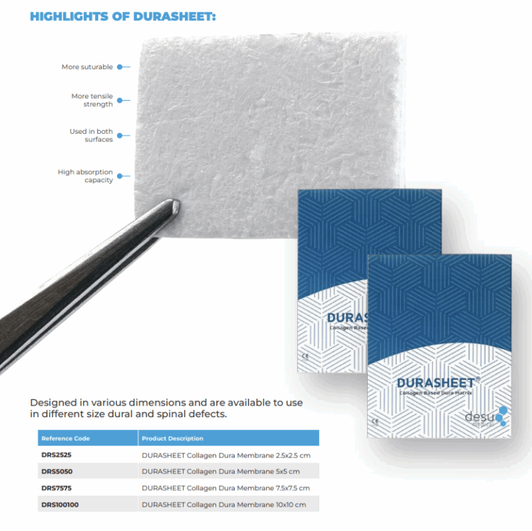
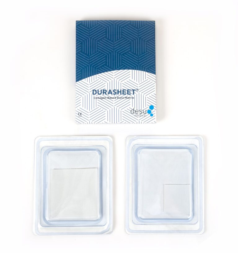
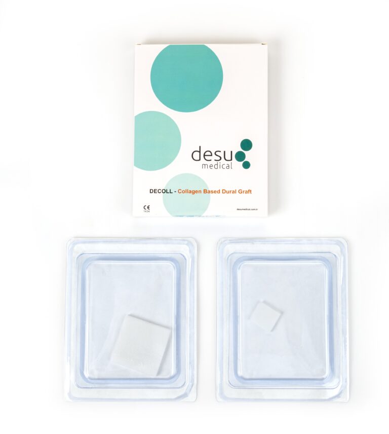
Autologous pericranium or fascia from the same incision is used by many surgeons, particularly in the midline approach. Natural collagen grafts are a common alternative because they are available in different sizes, can be trimmed to irregular shapes and are gradually replaced by the patient’s own tissue. Desu’s DURASHEET and DECOLL are made from natural, pure Type I bovine collagen with an impermeable pore structure designed to act as a barrier against CSF. They are supplied sterile and ready to use, with no pre-treatment.
Bone Flap or Craniectomy?
Some posterior fossa operations are performed as a craniectomy, in which bone is removed and not replaced, and others as a craniotomy, with the bone flap replaced at the end. Replacing the bone adds an extra layer over the dural repair and may reduce the risk of pseudomeningocele, but practice varies with the approach and the surgeon’s preference.
Managing Hydrocephalus
Tumours of the posterior fossa often obstruct CSF flow and cause hydrocephalus. If hydrocephalus persists after surgery, the raised CSF pressure stresses the dural closure and increases the risk of leakage and pseudomeningocele. Temporary external drainage, endoscopic third ventriculostomy or a shunt may be required. Desu manufactures external ventricular drainage systems and hydrocephalus shunts used in these situations.
After Surgery
Patients are observed for wound leakage, swelling under the incision, clear nasal discharge and signs of meningitis. A soft swelling may be a pseudomeningocele, which often settles with time but needs review if it grows or leaks. Clear fluid from the nose after a lateral approach suggests CSF rhinorrhoea through the mastoid air cells and requires prompt assessment.
Summary
Dural closure after posterior fossa surgery has to cope with retracted dura, nearby venous sinuses, high CSF pressure and a deep soft tissue bed. A tested dural repair, a graft where primary closure is not possible, sealing of mastoid air cells, a careful layered closure and management of hydrocephalus together reduce the risk of CSF leakage.
Frequently Asked Questions
Why are CSF leaks more common after posterior fossa surgery?
The dura in this region often retracts, venous sinuses limit suturing, CSF pressure can be high, and the deep muscle layers leave room for fluid to collect. Lateral approaches may also open mastoid air cells.
Is a dural graft always needed after posterior fossa surgery?
No. When the edges can be closed without tension, primary closure is used. A graft is needed when the dura cannot be closed directly or when an expansile duraplasty is planned.
What is CSF rhinorrhoea after posterior fossa surgery?
It is leakage of cerebrospinal fluid through the nose, which can occur after lateral approaches if CSF tracks through opened mastoid air cells to the middle ear and Eustachian tube.
References and Further Reading
- PubMed: CSF leak after posterior fossa surgery
- PubMed: retrosigmoid approach and CSF leak
- Desu Medical, DURASHEET collagen dura membrane
About this article
Prepared by the Desu Medical editorial team. Desu Medical has designed and manufactured neurosurgical devices in Ankara, Türkiye, since 2007, including the DURASHEET and DECOLL collagen dura membranes, which are made from natural, pure Type I bovine collagen. Manufacturing operates under an ISO 13485 quality management system and the dural graft range is CE certified. This article is written for healthcare professionals and general information; it does not replace the Instructions for Use or clinical judgement. Last reviewed: October 2026.
