
Meningiomas are the most common primary tumours of the meninges, and most of them are attached to the dura mater. During meningioma resection, removing the tumour together with its dural attachment is one of the main ways to reduce the chance of recurrence. That leaves a dural defect, and repairing it is an essential part of the operation. This article explains why the dura is removed, how the defect is planned and which graft and closure options surgeons use.
Why the Dura Matters in Meningioma Resection
Meningiomas arise from cells of the arachnoid layer and typically grow from a base on the inner surface of the dura. On MRI, the adjacent dura often shows a thickened, enhancing strip known as the dural tail. Tumour cells can extend into the dura around the main attachment and, in some cases, into the overlying bone, which may become thickened (hyperostosis).
The extent of resection is classically described with the Simpson grading system:
- Grade I: complete removal of the tumour, its dural attachment and any abnormal bone.
- Grade II: complete removal of the tumour with coagulation of the dural attachment.
- Grade III: complete removal of the tumour without resection or coagulation of the dural attachment.
- Grade IV: partial removal.
- Grade V: decompression or biopsy only.
Higher grades have historically been associated with higher recurrence rates, which is why surgeons aim to remove the involved dura when it is safe to do so. A Simpson grade I meningioma resection therefore almost always requires a duraplasty.
Planning the Dural Defect
The size and shape of the dural defect are largely known before surgery from MRI. Surgeons typically remove the dura with a margin of normal-appearing tissue around the attachment. Planning considers:
- the location of the tumour, for example convexity, parasagittal, falcine or skull base;
- involvement of venous sinuses, which may limit how much dura can be removed;
- bone involvement and the need for cranioplasty;
- the condition of the scalp and any previous surgery or radiotherapy.
Graft Options After Meningioma Resection
For small defects, autologous pericranium or temporalis fascia may be available in the operative field. For larger convexity defects, or when the surgeon prefers to avoid additional harvesting, biological grafts of animal origin are widely used. Natural collagen grafts made from Type I bovine collagen are popular because they come in several sizes, can be trimmed to irregular shapes and are gradually remodelled into the patient’s own tissue.
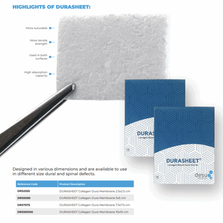
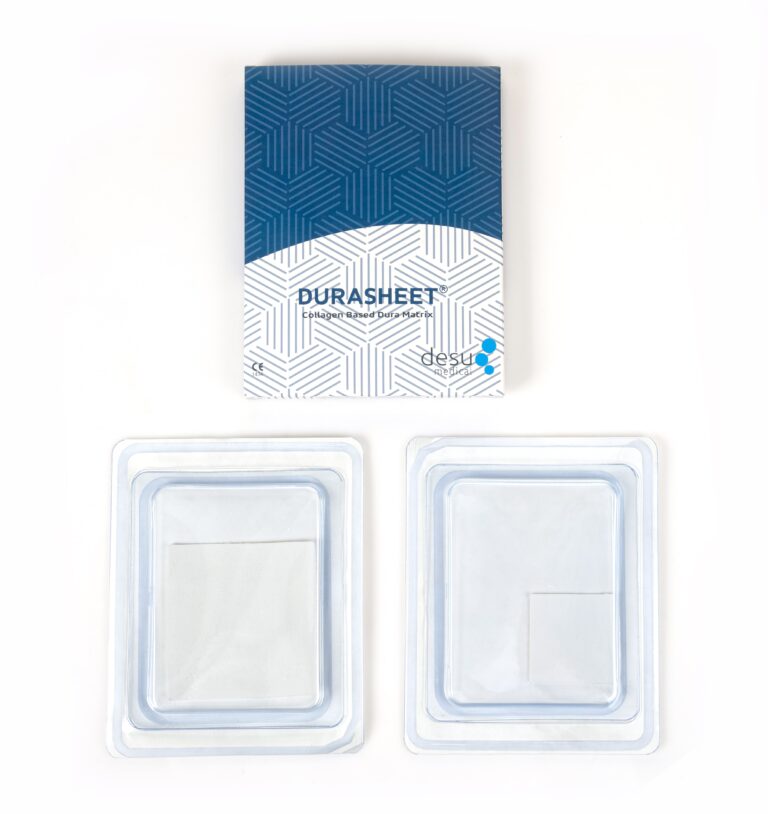
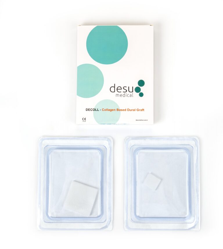
Desu’s DURASHEET and DECOLL membranes are made from natural, pure Type I bovine collagen and are supplied ready to use, so they can be cut to the shape of the defect directly at the table.
Closure Techniques
Convexity Meningiomas
Convexity defects are usually accessible on all sides, which allows the graft to be sutured circumferentially to the dural edges or placed as an onlay with an overlap under the dural margins. A watertight closure, tested with a Valsalva manoeuvre, reduces the risk of subgaleal CSF collection.
Parasagittal and Falcine Meningiomas
Near the superior sagittal sinus, the dural edge may be incomplete on one side. The graft is then tucked under the remaining dura or secured to the falx where possible, and the repair is often reinforced with an adjunct.
Skull Base Meningiomas
At the skull base, suturing may be difficult or impossible. Multilayer reconstruction, using inlay and onlay grafts combined with vascularised tissue where available, is common. Our article on skull base dural repair covers these techniques.
Recurrent Tumours and Irradiated Fields
Repeat meningioma resection, or surgery after radiotherapy, brings additional challenges. Scar tissue can make the dural edges thin and fragile, the scalp may heal more slowly, and the remaining dura may not hold sutures well. In these settings surgeons often plan a generous graft with a wide overlap, avoid tension on the dural edges and pay particular attention to the layered closure of the scalp. A graft that conforms easily to the surface and integrates into surrounding tissue is helpful when the local tissue quality is poor. Close follow-up of the wound is advisable after a repeat meningioma resection.
After Meningioma Resection
Postoperative care focuses on wound healing, detection of CSF collections and monitoring for infection. MRI at follow-up assesses the extent of resection and any residual tumour. A well-integrated collagen graft is not expected to cause long-term imaging problems, although early postoperative images may show enhancement and fluid along the operative bed as part of normal healing.
Summary
A thorough meningioma resection frequently means removing the dural attachment, and a careful duraplasty completes the operation. Planning the defect from imaging, choosing a graft that can be shaped to fit and achieving a watertight closure help protect the patient from CSF leakage and wound complications.
Frequently Asked Questions
Why is the dura removed during meningioma resection?
Meningiomas grow from the dura and tumour cells can extend into it. Removing the dural attachment, described as Simpson grade I, is associated with a lower chance of recurrence than leaving it in place.
What graft is used after meningioma resection?
Autologous tissue such as pericranium can be used for small defects. For larger defects many surgeons use natural collagen grafts made from Type I bovine collagen, which can be trimmed to shape and integrate into host tissue.
Is the dura always repaired with a graft?
Not always. If only a small strip of dura is removed, the edges may be closed directly. A graft is needed when the defect cannot be closed without tension.
References and Further Reading
- PubMed: Simpson grade and meningioma recurrence
- PubMed: duraplasty after meningioma resection
- Desu Medical, Dural Grafts product information
About this article
Prepared by the Desu Medical editorial team. Desu Medical has designed and manufactured neurosurgical devices in Ankara, Türkiye, since 2007, including the DURASHEET and DECOLL collagen dura membranes, which are made from natural, pure Type I bovine collagen. Manufacturing operates under an ISO 13485 quality management system and the dural graft range is CE certified. This article is written for healthcare professionals and general information; it does not replace the Instructions for Use or clinical judgement. Last reviewed: October 2026.
