
Operations at the skull base pose a particular challenge for dural repair. Whether the approach is endoscopic through the nose or open through the skull, the dural defect often lies in a narrow corridor, against bone, where conventional suturing is difficult or impossible. Yet a reliable repair is essential, because a cerebrospinal fluid (CSF) leak at the skull base can lead to meningitis. This article explains the principles of skull base reconstruction and where collagen dural grafts fit in.
Why Skull Base Defects Are Different
At the convexity, a surgeon can usually suture a graft to the dural edges on all sides. At the skull base, the dura is closely attached to irregular bone, the defect may be surrounded by critical neurovascular structures, and the repair has to withstand CSF pulsation from above and, after endonasal surgery, contact with the sinonasal cavity below. The aim is therefore not a single watertight suture line but a layered barrier that seals the defect while healing takes place.
Endoscopic Endonasal Skull Base Surgery
Endoscopic endonasal approaches are used for pituitary adenomas, craniopharyngiomas, clival chordomas, some anterior skull base meningiomas and other lesions. These operations can create defects ranging from a small leak through the diaphragma sellae to a large opening of the anterior skull base with high CSF flow. The reconstruction is chosen according to the size of the defect and the expected flow of CSF.
Multilayer Reconstruction
Most teams build the repair in layers:
- Inlay graft: placed inside the defect, between the dura and the bone or in the subdural space, to act as the first barrier.
- Onlay graft: placed over the bony defect on the nasal side, overlapping the edges.
- Vascularised flap: commonly the nasoseptal flap, which brings its own blood supply and is particularly valuable for large, high-flow defects.
- Supporting materials: fat grafts, sealants and nasal packing that hold the layers in place during early healing.
Collagen dural grafts are widely used as inlay or onlay layers because they are soft, conform to irregular surfaces and are resorbed as the patient’s own tissue grows into them.
Small Sellar Defects
Not every endonasal operation produces a large opening. After many pituitary operations the leak is small or there is only a weeping of CSF through a thinned diaphragma sellae. In these cases a simpler repair is often enough: a small piece of collagen graft placed as an inlay or onlay, sometimes supported by a fat graft or sealant. Matching the extent of the reconstruction to the size of the defect avoids unnecessary morbidity, such as harvesting a nasoseptal flap when it is not needed.
Open Skull Base Approaches
Open approaches to the anterior and middle cranial fossa, for example for olfactory groove meningiomas or tumours invading the floor of the skull, also require careful dural repair. Surgeons frequently combine a dural graft with a vascularised pericranial flap that is turned down over the floor of the anterior cranial fossa to separate the intracranial space from the paranasal sinuses. Our article on dural repair after meningioma resection discusses tumour-related defects in more detail.
Choosing a Graft for Skull Base Reconstruction
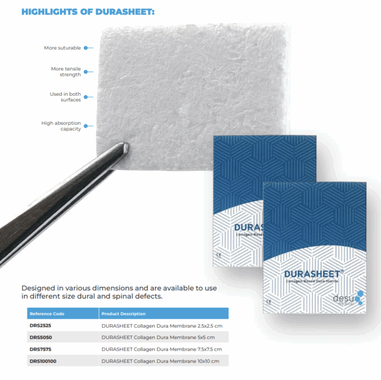
The ideal graft for skull base work is easy to cut to small, irregular shapes, handles well through a narrow corridor, adheres to the surface without migrating and integrates into host tissue. Natural collagen grafts made from Type I bovine collagen meet many of these needs. They are supplied ready to use, can be trimmed at the table and do not require suturing when used as an inlay or onlay layer.
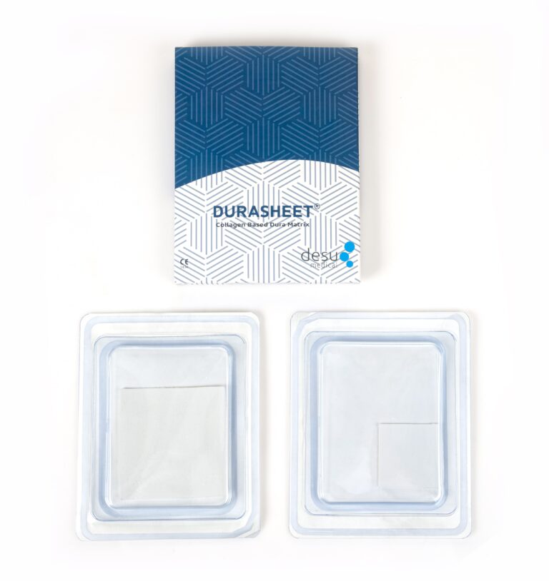
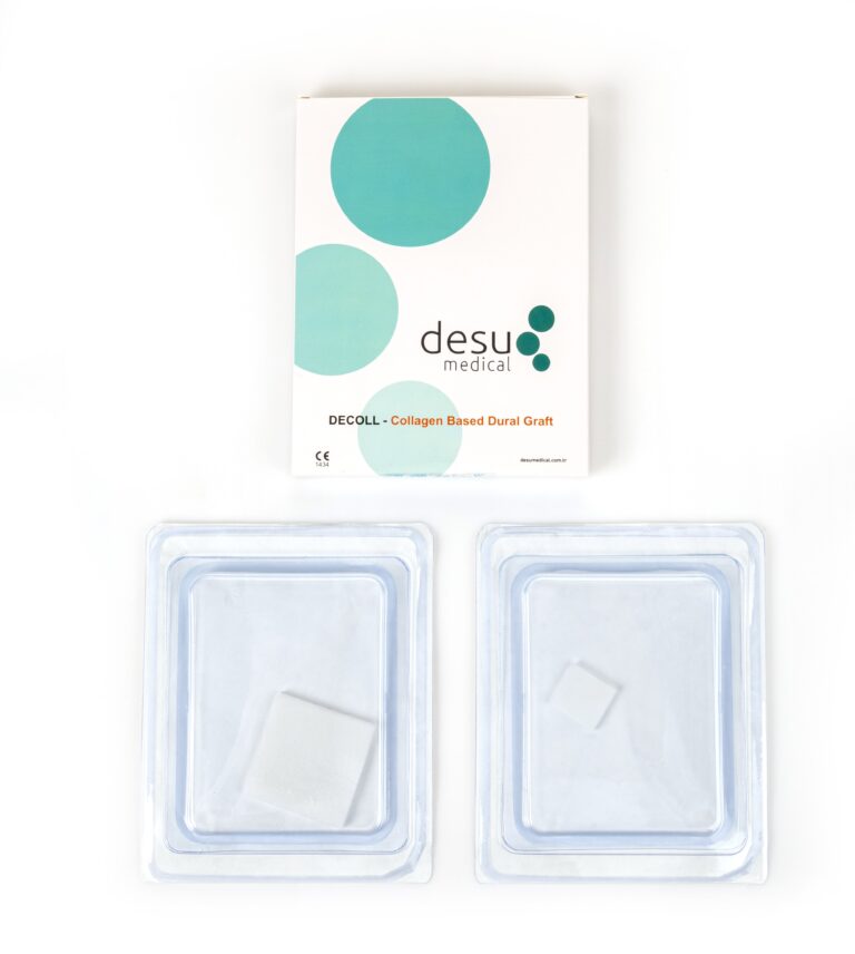
Desu’s DECOLL and DURASHEET membranes are made from natural, pure Type I bovine collagen and have an impermeable pore structure designed to act as a barrier against CSF. The smaller DECOLL formats are convenient for sellar and focal anterior skull base defects.
Managing High-Flow CSF Leaks
When a large cistern or the third ventricle has been opened, CSF flow against the repair is high. In these cases surgeons rely more heavily on vascularised tissue, robust multilayer reconstruction and, in selected patients, temporary CSF diversion with a lumbar drain. The skull base repair is only one part of a strategy that also includes careful patient selection and postoperative precautions.
Postoperative Care
After skull base surgery, patients are commonly advised to avoid nose blowing, straining and heavy lifting for a period, and to report clear nasal discharge, especially if it increases when bending forward. Early recognition of a CSF leak allows prompt management, which may include imaging, a lumbar drain or surgical revision. Signs of meningitis, such as fever, headache and neck stiffness, require urgent assessment.
Summary
Skull base reconstruction depends on layering: a well-placed inlay, an overlapping onlay, vascularised tissue where needed and supportive measures during healing. Natural collagen grafts contribute to this strategy by providing a conformable, resorbable barrier that becomes part of the patient’s own repair.
Frequently Asked Questions
Why is a CSF leak at the skull base dangerous?
After endonasal or anterior skull base surgery, a leak connects the space around the brain with the nose and sinuses, which carries a risk of meningitis. This is why skull base reconstruction is built in several layers.
Are collagen grafts sutured at the skull base?
Usually not. At the skull base collagen grafts are typically used as inlay or onlay layers held in place by other layers, a vascularised flap, sealant or packing.
What is a nasoseptal flap?
It is a flap of the nasal septum lining with its own blood supply, rotated to cover the skull base defect after endonasal surgery. It is often used together with dural grafts for large defects.
References and Further Reading
- PubMed: endoscopic skull base reconstruction
- PubMed: nasoseptal flap
- Desu Medical, DECOLL collagen dura membrane
About this article
Prepared by the Desu Medical editorial team. Desu Medical has designed and manufactured neurosurgical devices in Ankara, Türkiye, since 2007, including the DURASHEET and DECOLL collagen dura membranes, which are made from natural, pure Type I bovine collagen. Manufacturing operates under an ISO 13485 quality management system and the dural graft range is CE certified. This article is written for healthcare professionals and general information; it does not replace the Instructions for Use or clinical judgement. Last reviewed: October 2026.

Comments (2)
Dural Repair Glossary: Clear Definitions of 35 Key Terms
[…] Skull base: the floor of the skull, on which the brain rests. See skull base dural repair. […]
Meningioma Resection: Clear Dural Repair Guide (5 Simpson Grades)
[…] inlay and onlay grafts combined with vascularised tissue where available, is common. Our article on skull base dural repair covers these […]