
The dura mater is the tough, outermost of the three membranes that surround the brain and spinal cord. Its name comes from Latin and means “tough mother”, a fair description of a layer that protects the central nervous system and keeps cerebrospinal fluid (CSF) where it belongs. When the dura mater is opened during surgery, damaged by trauma or removed together with a tumour, restoring it becomes an important step in protecting the patient from CSF leakage, infection and wound problems.
This guide explains how the dura mater is built, what it does, and how neurosurgeons repair it when it cannot simply be sutured closed.
Key Points
- The dura mater is the outer layer of the meninges, above the arachnoid and pia mater.
- Cranial dura has two layers; between them run the dural venous sinuses.
- Spinal dura forms a single tube, the dural sac, which holds CSF around the spinal cord and nerve roots.
- The dura mater is dense connective tissue rich in Type I collagen, which is why collagen-based grafts are biologically familiar to it.
The Three Meninges
The brain and spinal cord are wrapped in three membranes, collectively called the meninges. From outside to inside they are the dura mater, the arachnoid mater and the pia mater. The arachnoid is a thin, delicate layer, and beneath it lies the subarachnoid space, where CSF circulates. The pia mater is the innermost layer and follows every fold of the brain and spinal cord surface.
Between the bone and the dura mater lies the epidural space. In the skull it is only a potential space, because the dura is firmly attached to the inner surface of the bone. In the spine it is a real space containing fat and a network of veins. Between the dura and the arachnoid lies the subdural space, which is also normally only a potential space.
Structure of the Cranial Dura Mater
Inside the skull the dura mater has two layers. The outer, periosteal layer lines the inner surface of the skull bones. The inner, meningeal layer is continuous with the spinal dura. For most of their extent the two layers are fused, but in certain places they separate to enclose the dural venous sinuses, the channels that drain venous blood from the brain.
The meningeal layer also folds inwards to form partitions that support the brain and limit its movement:
- Falx cerebri: a sickle-shaped fold between the two cerebral hemispheres.
- Tentorium cerebelli: a tent-like sheet separating the cerebrum from the cerebellum.
- Falx cerebelli: a small fold between the cerebellar hemispheres.
- Diaphragma sellae: a roof over the pituitary fossa, pierced by the pituitary stalk.
The Spinal Dura Mater
In the spine the dura mater forms a single-layered tube, often called the dural sac or thecal sac. It begins at the foramen magnum, where it is continuous with the cranial dura, and usually ends at about the level of the second sacral vertebra. Along its length it sends sleeves around the spinal nerve roots as they exit through the intervertebral foramina.
Because the spinal dural sac is a closed, fluid-filled tube, even a small opening can allow CSF to escape. This is why incidental durotomy during spine surgery receives careful attention, and why our article on spinal dural repair focuses on watertight closure.
What the Dura Mater Does
The dura mater has several functions that explain why its integrity matters after surgery:
- Mechanical protection: it is the strongest of the meninges and forms a durable envelope around neural tissue.
- Containing CSF: together with the arachnoid, it keeps CSF within the subarachnoid space, where it cushions the brain and spinal cord.
- Venous drainage: the dural venous sinuses collect blood from the brain and return it towards the internal jugular veins.
- Barrier function: an intact dura separates the central nervous system from the outside environment, which matters greatly after an operation.
- Sensation: the dura is innervated, mainly by branches of the trigeminal nerve and upper cervical nerves, which is one reason why some headaches originate from dural structures.
What the Dura Mater Is Made Of
Histologically, the dura mater is dense irregular connective tissue. Its strength comes from bundles of collagen fibres, predominantly Type I collagen, arranged in interwoven layers, with fibroblasts and a smaller amount of elastic fibres between them. This composition has a practical consequence for dural repair: a graft built from Type I collagen presents surrounding cells with a protein they already recognise. You can read more about this in our article on Type I collagen and natural dural grafts.
When the Dura Mater Needs Repair
Many operations involve opening the dura mater, and in most cases the surgeon closes it again with sutures. A dural graft becomes necessary when the edges cannot be brought together without tension, or when part of the dura has been removed. Typical situations include:
- resection of tumours attached to or invading the dura, such as meningiomas;
- decompressive procedures, for example expansile duraplasty in Chiari malformation or after decompressive craniectomy;
- traumatic tears with tissue loss;
- dural defects in spine surgery, including intradural tumour removal;
- repair of congenital defects in children.
Surgeons can choose between autologous tissue such as pericranium or fascia, biological grafts of animal origin, and synthetic materials. Each option involves trade-offs in availability, handling, integration and long-term behaviour.
How a Natural Collagen Graft Supports Dural Repair
A collagen dural graft acts as a scaffold. After implantation, fibroblasts from the surrounding tissue migrate into the matrix and begin laying down new connective tissue. Over time the graft is gradually resorbed and replaced by the patient’s own tissue, often described as a neodura. Because the graft does not remain as a permanent foreign body, it avoids some of the long-term concerns associated with non-resorbable synthetic substitutes.
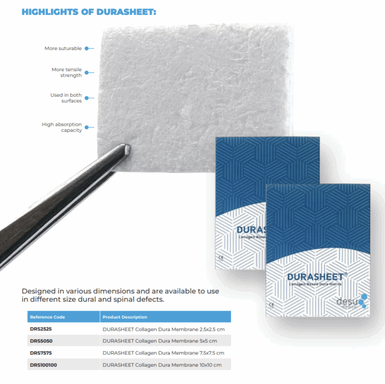
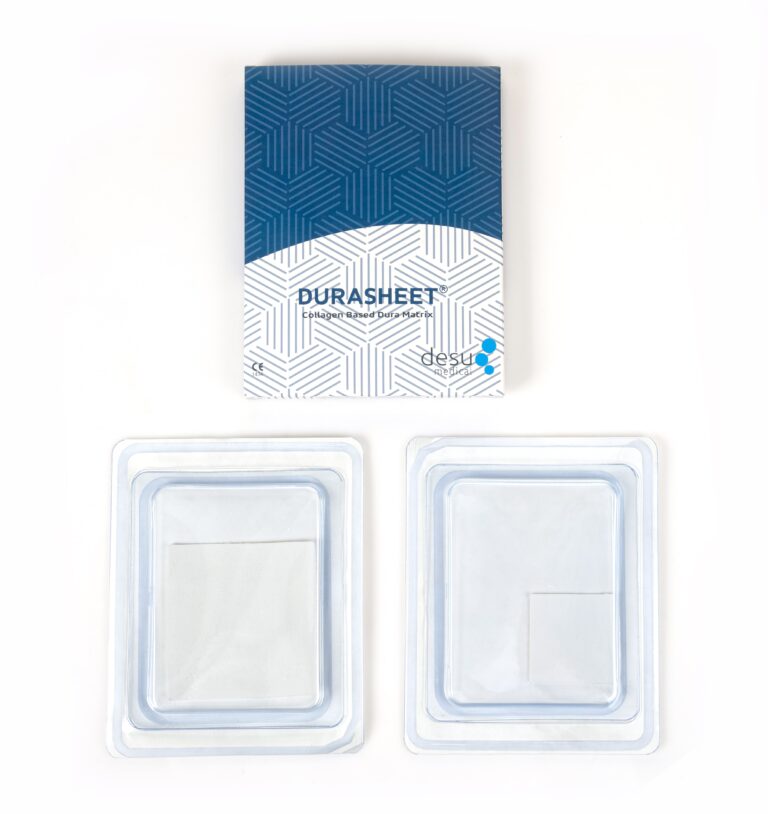
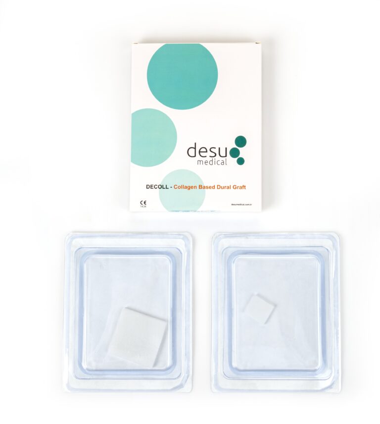
Desu manufactures its DURASHEET and DECOLL collagen dura membranes from natural, pure Type I bovine collagen, not from synthetic polymers. Both are single-use, gamma sterilised and ready to use, with an impermeable pore structure designed to act as a barrier against CSF leakage while the dura mater heals.
Frequently Asked Questions
Is the dura mater the same as the meninges?
No. The meninges are three membranes: the dura mater, the arachnoid mater and the pia mater. The dura mater is the outermost and strongest of the three.
What happens if the dura mater is damaged?
A breach in the dura mater can allow cerebrospinal fluid to leak, which may lead to a fluid collection, headache, wound problems or, in some cases, infection. This is why surgeons aim for a watertight dural closure.
Can the dura mater heal on its own?
Small, well-apposed openings can heal after suturing. Larger defects usually need a graft that bridges the gap and provides a scaffold for new tissue, which over time becomes part of the patient’s own dura mater.
References and Further Reading
- PubMed: dura mater anatomy
- PubMed: duraplasty and dural substitutes
- Desu Medical, Dural Grafts product information
About this article
Prepared by the Desu Medical editorial team. Desu Medical has designed and manufactured neurosurgical devices in Ankara, Türkiye, since 2007, including the DURASHEET and DECOLL collagen dura membranes, which are made from natural, pure Type I bovine collagen. Manufacturing operates under an ISO 13485 quality management system and the dural graft range is CE certified. This article is written for healthcare professionals and general information; it does not replace the Instructions for Use or clinical judgement. Last reviewed: October 2026.
