
Removing an intradural spinal tumour requires opening the dura of the spinal canal. Schwannomas, meningiomas, ependymomas and other lesions inside the dural sac can only be reached through this opening, and the operation is not complete until the dura has been closed again. A reliable closure protects the patient from CSF leakage, pseudomeningocele and infection. This article describes how surgeons approach dural closure after these operations.
Types of Intradural Spinal Tumour
Intradural tumours are divided into two groups. Intradural extramedullary tumours lie inside the dura but outside the spinal cord; schwannomas, neurofibromas and meningiomas are the most common examples. Intramedullary tumours, such as ependymomas and astrocytomas, grow within the spinal cord itself. In both cases the dura is opened in the midline, usually over the full length of the tumour, and held open with stay sutures while the tumour is removed under the microscope.
Why Spinal Dural Closure Is Demanding
CSF pressure in the spinal canal rises when the patient sits or stands, and the lumbar region in particular carries a high hydrostatic load. The dural closure must withstand this pressure from the first day. At the same time, the spinal dural sac is narrow, the surgical field is deep and the closure is often performed through a limited exposure. Leaks after spinal surgery tend to track into the muscle layers and can form a pseudomeningocele or a cutaneous fistula.
Primary Closure
In most cases of intradural spinal tumour surgery, the dural edges can be brought together directly with a running or interrupted fine suture. Primary closure is the preferred option when the edges are healthy and there is no tension. The surgeon then tests the repair, often by asking the anaesthetist to perform a Valsalva manoeuvre, and reinforces any weak points.
When a Graft Is Needed
A dural graft is used when primary closure is not possible or would narrow the dural sac. Typical situations include:
- Dural meningiomas: where the dural attachment of the tumour is removed with it, leaving a defect.
- Tumours with dural infiltration: where part of the dura must be excised.
- Swollen spinal cord: after resection of an intramedullary tumour, when a patch enlarges the dural sac and avoids compression.
- Thin or torn edges: that will not hold sutures.
Closure Technique in 5 Steps
- Prepare the edges: trim irregular margins and identify healthy dura.
- Size the graft: cut the graft slightly larger than the defect so that it overlaps the edges.
- Secure the graft: suture it with a fine running suture or place it according to the chosen onlay technique.
- Test the repair: perform a Valsalva manoeuvre and close any leaking points.
- Close in layers: close muscle, fascia and skin carefully, avoiding dead space above the dura.
Graft Choice
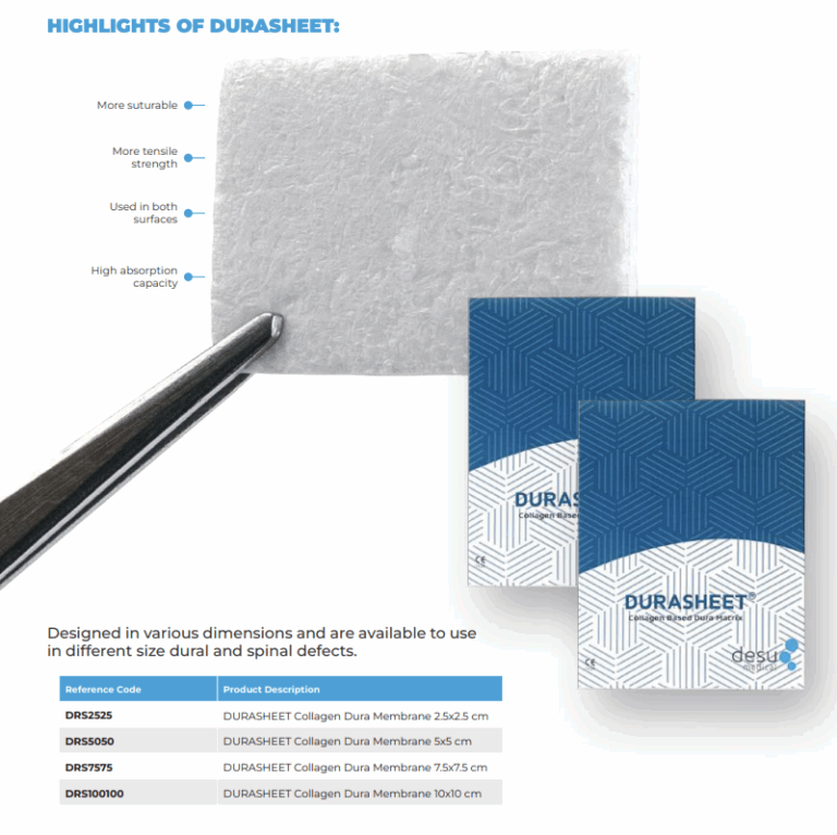
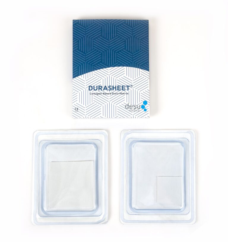
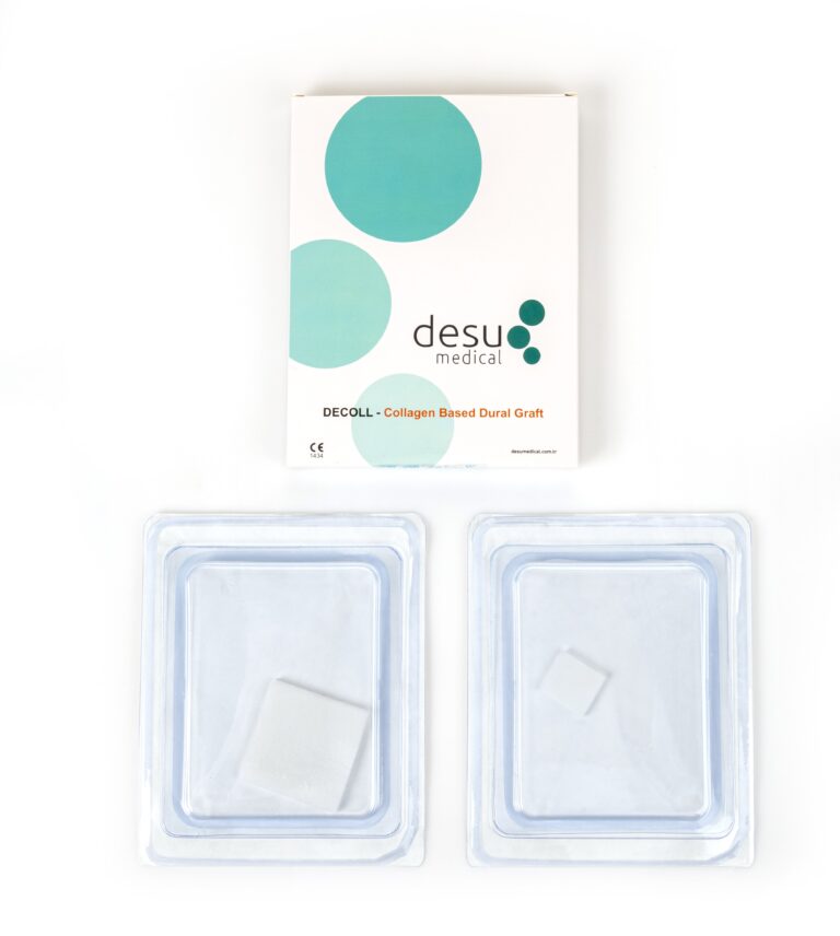
The choice of graft affects how well the closure performs after intradural spinal tumour removal. For intradural spinal tumour surgery, surgeons look for a graft that is thin enough to lie flat in a narrow canal, strong enough to hold sutures and able to integrate with the surrounding tissue. Natural collagen grafts made from Type I bovine collagen meet these requirements and are gradually replaced by the patient’s own connective tissue. Desu’s DURASHEET and DECOLL membranes are supplied sterile and ready to use, and the DECOLL range includes small formats suited to focal spinal defects. Our article on spinal dural repair covers incidental durotomy, a related situation.
Intramedullary Tumours and the Swollen Cord
After removal of an intramedullary intradural spinal tumour, the spinal cord can be swollen. Closing the dura tightly over a swollen cord may compress it. Some surgeons therefore use an expansile graft to enlarge the dural sac, in the same way as an expansile duraplasty in the posterior fossa. The graft is sutured into the dural opening so that the cord has room while the swelling settles.
Adjuncts
Sealants may be applied over the suture line to reinforce it. Fat or muscle grafts can fill dead space above the dura. Lumbar drainage is sometimes used for a short period in high-risk cases to reduce CSF pressure on the repair, although many surgeons reserve it for leaks that appear after surgery.
Postoperative Care
Postoperative routines after intradural spinal tumour resection differ between centres. Practices vary, but some teams keep patients flat for a period after intradural spinal tumour surgery, particularly after lumbar procedures, to reduce pressure on the closure. Patients are monitored for wound leakage, positional headache and swelling under the incision. Persistent leakage requires prompt assessment because of the risk of meningitis.
Summary
Dural closure is the final, essential step of intradural spinal tumour surgery. Primary closure is preferred when possible, while a dural graft is used for defects, infiltrated dura or when the sac must be enlarged. A tested repair and a careful layered soft tissue closure protect the result.
Frequently Asked Questions
Is a dural graft always needed after intradural spinal tumour surgery?
No. Most closures are performed by suturing the dura directly. A graft is used when there is a defect, when dura has been excised with the tumour or when the dural sac must be enlarged.
Why are patients sometimes kept flat after spinal tumour surgery?
Lying flat lowers CSF pressure in the lumbar region and reduces the load on the dural closure during early healing. Practices differ between surgical teams.
What happens if the dural closure leaks?
A leak may cause a fluid collection, positional headache or leakage through the wound. Treatment ranges from observation and drainage to surgical repair.
References and Further Reading
- PubMed: intradural spinal tumour dural closure
- PubMed: spinal meningioma dural resection
- Desu Medical, DURASHEET collagen dura membrane
About this article
Prepared by the Desu Medical editorial team. Desu Medical has designed and manufactured neurosurgical devices in Ankara, Türkiye, since 2007, including the DURASHEET and DECOLL collagen dura membranes, which are made from natural, pure Type I bovine collagen. Manufacturing operates under an ISO 13485 quality management system and the dural graft range is CE certified. This article is written for healthcare professionals and general information; it does not replace the Instructions for Use or clinical judgement. Last reviewed: October 2026.
